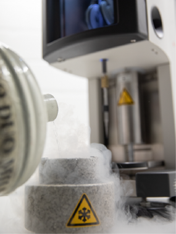
The Transmission Electron Microscopy Laboratory provides the scientific community with structural and spectroscopic analysis techniques with atomic spatial resolution. For this purpose, it is equipped with four transmission electron microscopes containing cameras for capturing images and electron diffraction and spectrometers for capturing X-ray photons characteristic of the sample, as well as for measuring the energy loss of the electrons after interacting with the sample. Thus, the size, morphology, crystalline structure, chemical composition, and electronic state (oxidation, reduction, hybridization, among others) of nanometric objects can be characterized with atomic resolution. In addition, the electrical and magnetic properties of the samples can be observed with nanometric resolution using the sample holders for heating liquids (23oC to 1200oC).
 Equipment
Thermo Fisher/FEI Titan Cubed Themis
Description: Transmission electron microscope double corrected and monochrome, with resolutions up to 0.6 Ångströms and two electron accelerating voltages (80 kV and 300 kV), to operate in TEM and STEM modes and analyze a variety of samples according to their sensitivity to electron beam. It has four EDS detectors to carry out high resolution X-ray spectroscopy mappings by energy dispersion, in a few minutes of collection. It has four different STEM detectors (HAADF, DF-conventional, DF-segmented and BF), to obtain high resolution images according to atomic number or by phase contrast. It also has a 4kx4k CMOS camera, with dynamic recording and two in situ sample holders: one for heating, to increase the temperature from ambient to 1300 °C, specific for samples of nanoparticles and coverslips prepared by FIB; and a liquid cell sample holder to analyze structures and dynamic processes of materials and biological samples in liquid environments, in static mode or in liquid flow.
Specifications:
Imaging Modes:
CTEM; HRTEM; BF-TEM; DF-TEM; NCSI-HRTEM (Negative Cs Imaging); BF-HRSTEM; ABF-HRSTEM; ADF-HRSTEM; HAADF-HRSTEM; DPC-HRSTEM; mono-HRTEM; mono-HRSTEM; Lorentz.
Electron Diffraction Modes:
SAED; NBD; CBED; PDF; 4D-STEM.
Spectroscopy Class(es):
EDS as atomic resolution.
Other Available Techniques:
TEM and STEM in situ heating for nanoparticles and coverslips by FIB; TEM and in situ STEM of liquid phases; TEM and STEM electron tomography; cryomicroscopy.

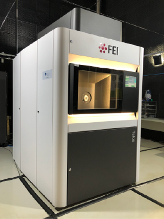
Talos F200C (Thermo Fisher Scientific)
Description: Transmission electron microscope with field-effect electron beam and accelerating voltage of 200 kV. Equipped with a 4k x 4k CMOS camera that allows low-dose acquisitions and a side-entry cryogenic sample holder. Software for automated image collection.
Specifications: Used for preliminary analyzes of structural biology, soft materials and electron diffraction.