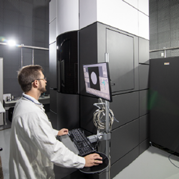
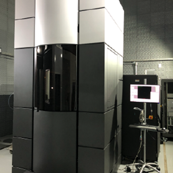
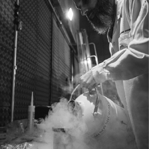
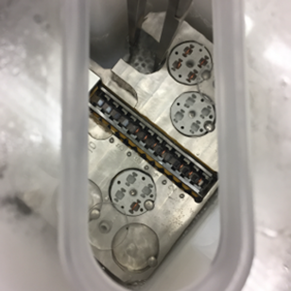
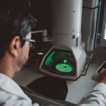
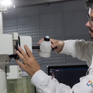
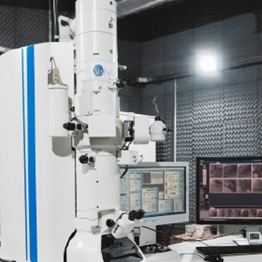
Cryo-ME (Cryogenic Transmission Electron Microscopy) is widely used to examine a range of systems, from soft materials to biomolecules, being a powerful technique for researching the structure of proteins. The immersion freezing method for vitrifying samples preserves the native structure of the particles, providing high-resolution structural information on biomolecules. LNNano has state-of-the-art transmission electron cryomicroscopes, two of which are fully equipped to support structural biology projects, with direct electron detectors, energy filters, and phase plates, operating at 200kV and 300kV. The sample preparation laboratories are equipped to support all the necessary sample preparation activities, including an NB-2 room.
 Equipments
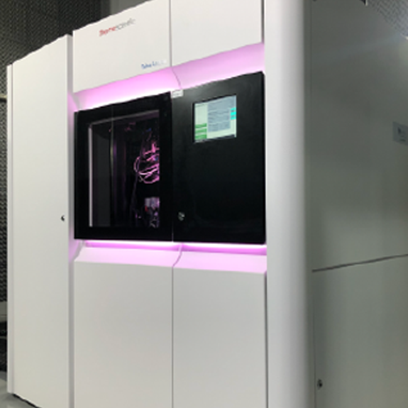
Titan Krios G3i (Thermo Fisher Scientific)
Specifications: Corrected transmission electron microscope with field-effect electron beam and accelerating voltage of 300 kV. Equipped with an automatic system for storage and exchange of grids, at cryogenic temperature. Equipped with a 4k x 4k CMOS camera, two direct electron detectors, one with an energy filter, and a “phase plate”. Software for automated image collection.
Techniques: Used for structural biology projects.
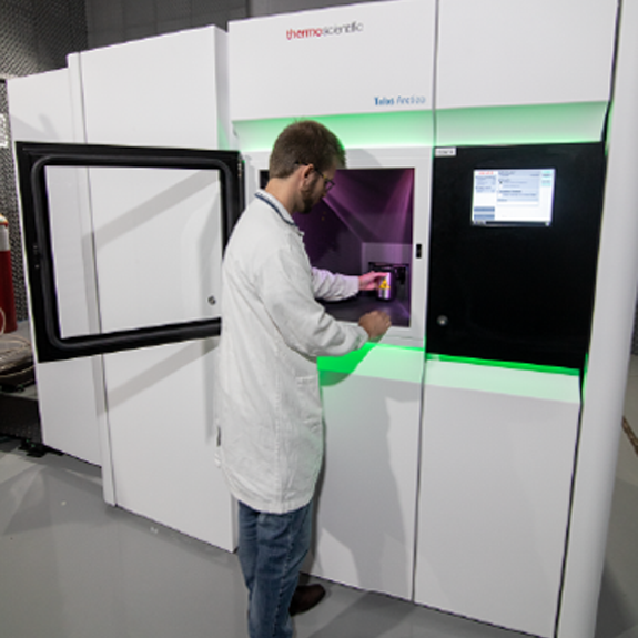
Talos Arctica G2 (Thermo Fisher Scientific)
Specifications: Transmission electron microscope with field-effect electron beam and accelerating voltage of 200 kV. Equipped with an automatic system for storage and exchange of grids, at cryogenic temperature. Equipped with a 4k x 4k CMOS camera, which allows for low-dose acquisitions, and a direct electron detector. Software for automated image collection.
Techniques: Used for optimizing structural biology projects and soft materials projects
JEM 1400 Plus (JEOL) - Porta amostras criogênico
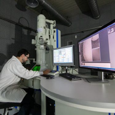
Specifications: Transmission electron microscope, with LaB6 electron gun and accelerating voltage of 120 kV. Equipped with a 4k x 4k CMOS camera that allows low-dose acquisitions and a side-entry cryogenic sample holder.
Techniques: Used for preliminary sample analysis, negative staining, cryoME and training.
- Get to know the division
- Facilities
- In-situ Growth Laboratory (LCIS)
- Spectroscopy and Scattering Laboratory
- Photoelectrochemistry Laboratory
- Transmission Electron Microscopy Laboratory
- Scanning and Dual-Beam Electron Microscopy Laboratory
- Atomic Force Microscopy Laboratory
- Nanoceramics Processing Laboratory
- Nanomaterials Synthesis Laboratory
- Staff
- Contact us


