Confocal Raman Spectrometer (Horiba, XploRA™ plus )
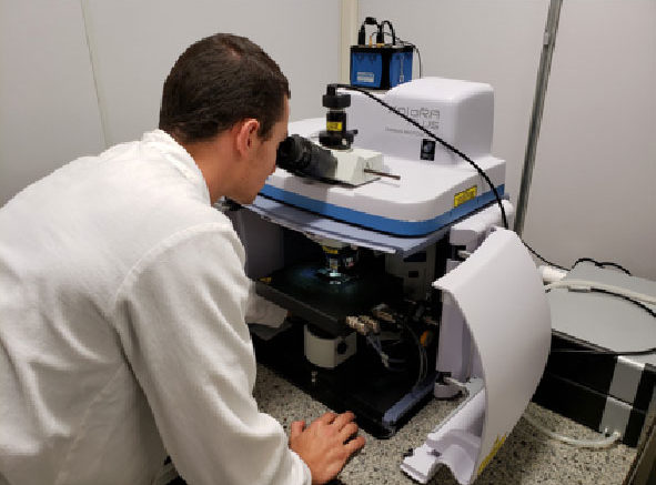
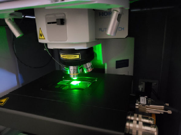
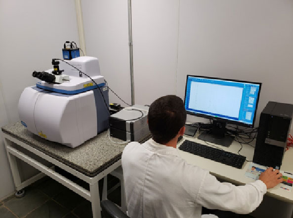
Confocal Raman Spectrometer (Horiba, XploRA™ plus )
[slick-slider design=”prodesign-6″ show_read_more=”false” dots=”false” arrows=”false” autoplay=”false” category=”696″ include_cat_child=”false” orderby=”title” order=”asc”]
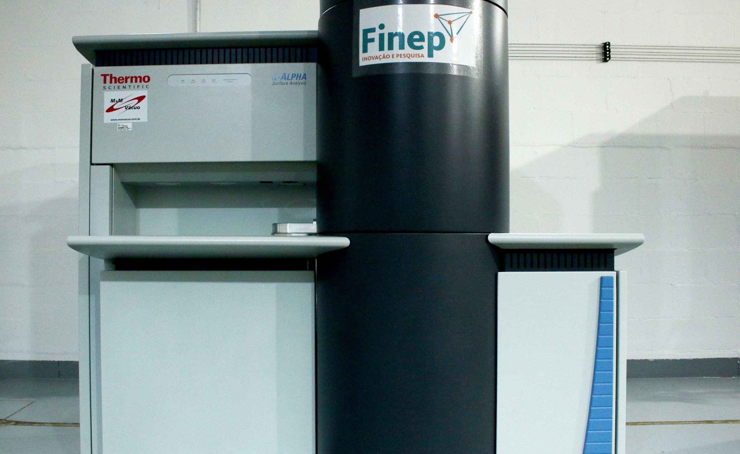
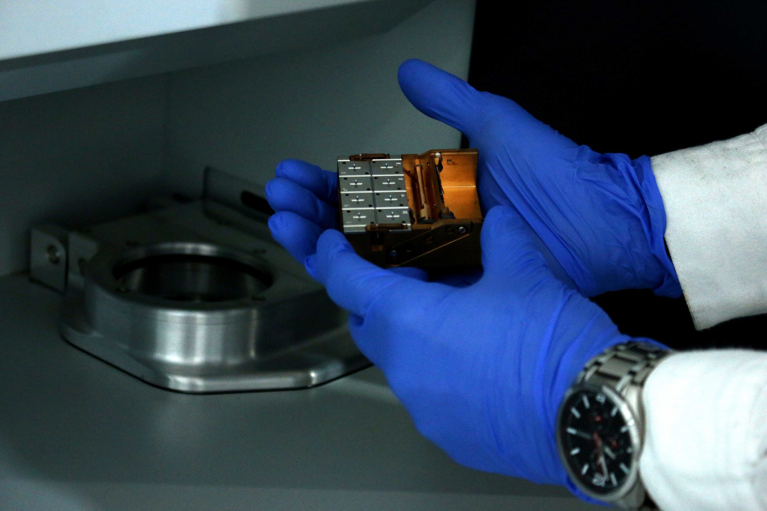
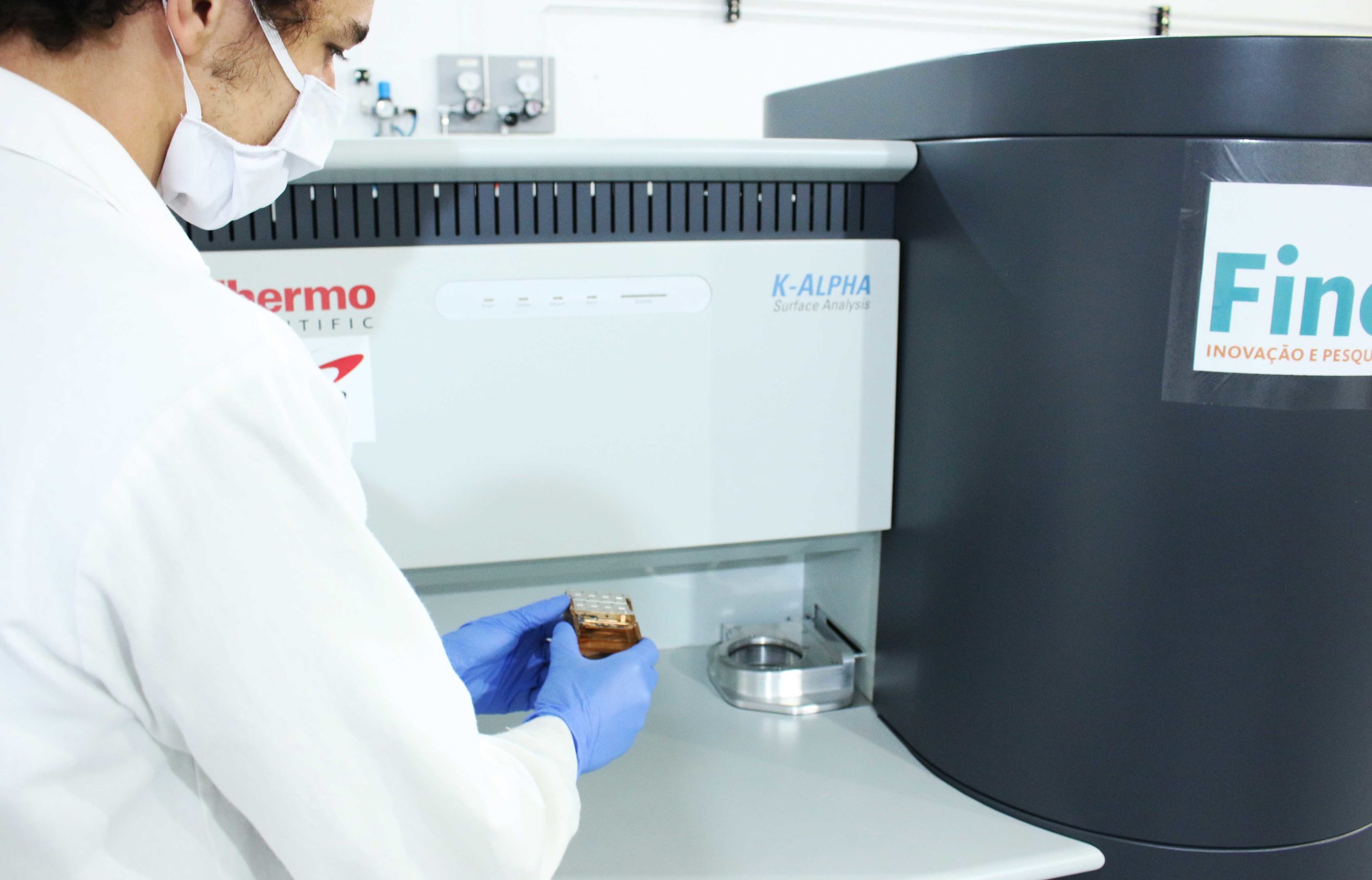
X-ray excited photoelectron spectrometer (XPS)
(Thermo Scientific, K-alpha)
X-ray excited photoelectron spectrometer (XPS)
(Thermo Scientific, K-alpha)
[slick-slider design=”prodesign-6″ show_read_more=”false” dots=”false” arrows=”false” autoplay=”false” category=”697″ include_cat_child=”false” orderby=”title” order=”asc”]
 SCATTERING
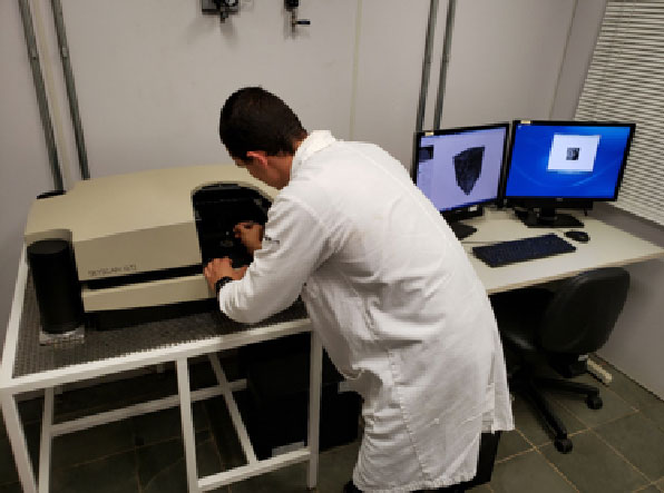
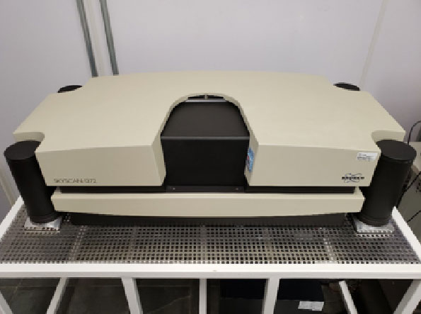
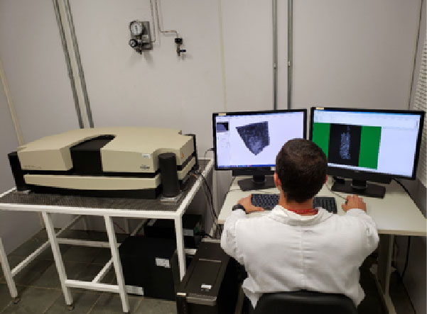
X-ray microtomography (Bruker (SkyScan) 1272)
X-ray microtomography (Bruker (SkyScan) 1272)
[slick-slider design=”prodesign-6″ show_read_more=”false” dots=”false” arrows=”false” autoplay=”false” category=”687″ include_cat_child=”false” orderby=”title” order=”asc”]
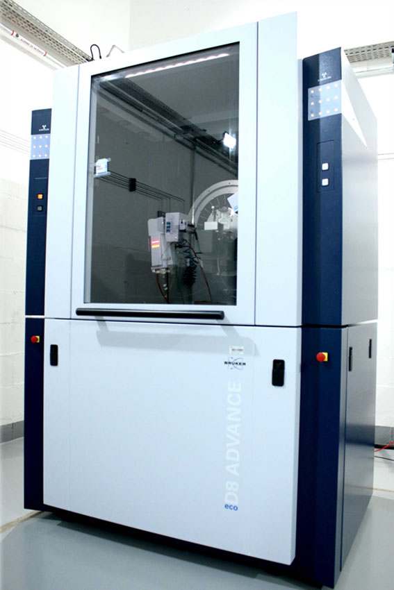
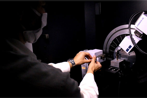
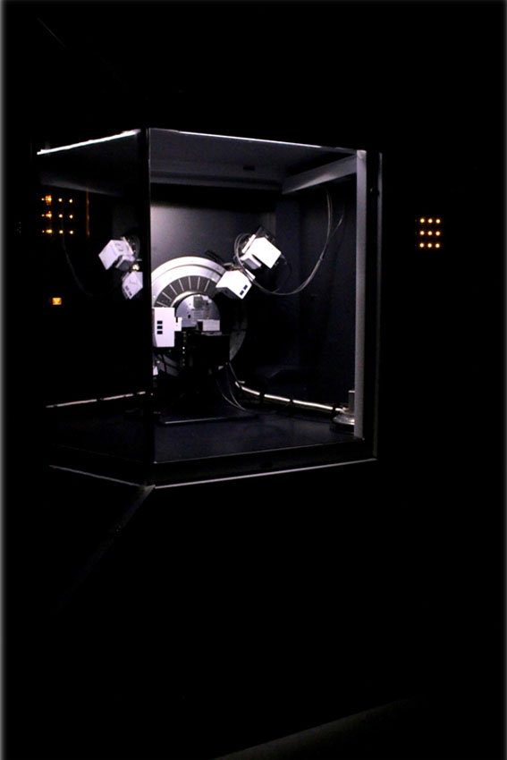
X-ray diffractometer (Bruker, D8 Advance Eco)