
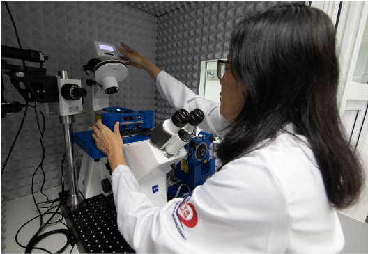
Atomic force microscopy (AFM) has become a very present tool in surface characterization, due to its operational flexibility, variety of analyzed properties and resolution at the nanometer scale.
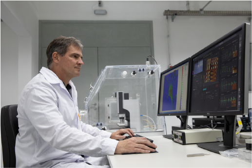
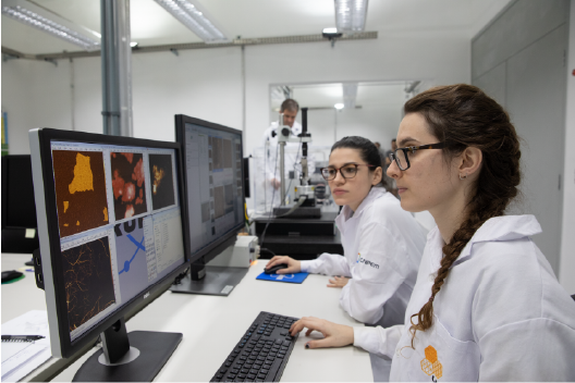
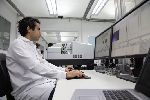
Open facilities have the mission of providing users with a research infrastructure with the best possible performance. They currently operate with four microscopes, with a wide variety of techniques related to the fields of physics, chemistry, materials science and biology. A team of specialists and technicians is available to assist in the execution of research projects, exploring the limits of available instruments.

Infrastructure
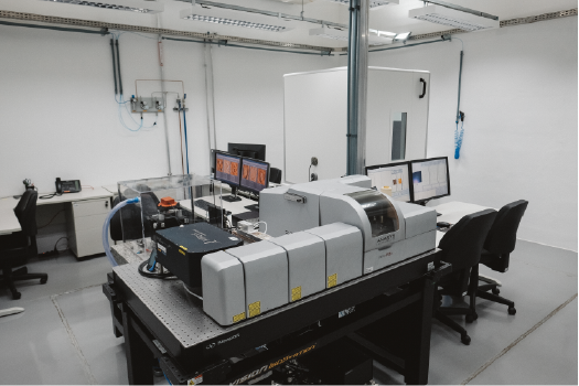
Park Systems - NX10
Park Systems - NX10
[slick-slider design=”prodesign-6″ show_read_more=”false” dots=”false” arrows=”false” autoplay=”false” category=”686″ include_cat_child=”false” orderby=”title” order=”asc”]
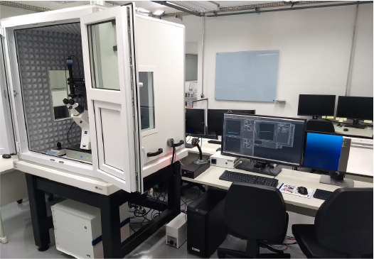
Bruker - MultiMode8
Bruker - MultiMode8
[slick-slider design=”prodesign-6″ show_read_more=”false” dots=”false” arrows=”false” autoplay=”false” category=”703″ include_cat_child=”false” orderby=”title” order=”asc”]
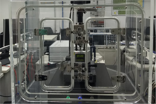
Bruker - NanoIR2-s
Bruker - NanoIR2-s
[slick-slider design=”prodesign-6″ show_read_more=”false” dots=”false” arrows=”false” autoplay=”false” category=”702″ include_cat_child=”false” orderby=”title” order=”asc”]
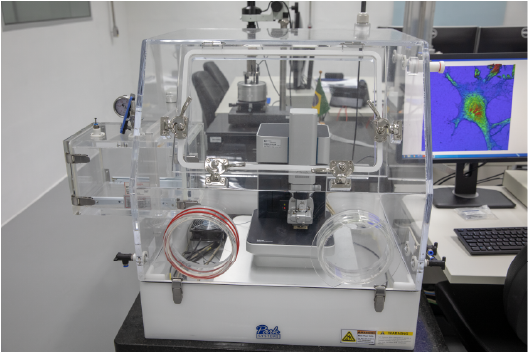
Bruker - BioAFM (NanoWizard4 e CellHesion200)
Bruker - BioAFM (NanoWizard4 e CellHesion200)
[slick-slider design=”prodesign-6″ show_read_more=”false” dots=”false” arrows=”false” autoplay=”false” category=”704″ include_cat_child=”false” orderby=”title” order=”asc”]