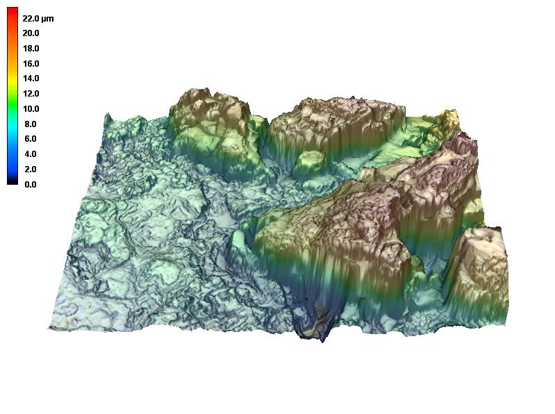
Confocal laser microscopy allows imaging and topology measurements with high depth of focus. The equipment combines conventional optical and laser microscopy to obtain images with high spatial resolution. These systems are known by the acronym LSM or LSCM (Laser Scanning Confocal Microscopy). Images are formed based on the light reflected by the sample. Through an embedded electronics it is possible to define the highest and lowest point (Z-axis) of the area of interest and slice the images so that several images of the same region are acquired, but with different points on the Z-axis. At the end of the measurement, the images are combined in such a way that it is possible to perform a three-dimensional (3D) reconstruction. The displacement step on the Z-axis can reach up to 10 nm. Analogous to the size of the probe by contact microscopy, the size of the optical “probe” basically depends on the wavelength used and the number of apertures of the lenses used to focus the light on the sample. In this system, the probe can reach up to 200 nm in diameter (x-y axis), for lenses with an NA of 0.95.
Equipment
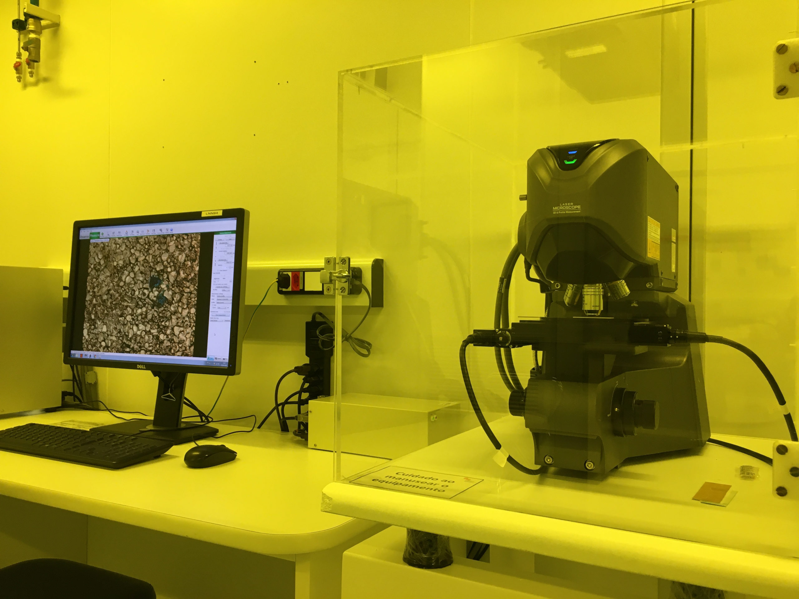
LSCM VK-X200 - Keyence
- Sample size: max. 100 x 100 x 28 mm (W x D x H);
Wavelength: 408 nm (violet);
Limited technique for transparent films smaller than 1 µm.
Available lenses:
| Lens | NA | NA lens working distance |
| 10x | 0.3 | 16.5 mm |
| 20x | 0.46 | 3.1 mm |
| 50x | 0.95 | 350 µm |
| 150x | 0.95 | 200 µm |
Equipment
LSCM VK-X200 - Keyence
-
- Sample size: max. 100 x 100 x 28 mm (W x D x H);
Wavelength: 408 nm (violet);
Limited technique for transparent films smaller than 1 µm.
Available lenses:
Lens NA NA lens working distance 10x 0.3 16.5 mm 20x 0.46 3.1 mm 50x 0.95 350 µm 150x 0.95 200 µm - Sample size: max. 100 x 100 x 28 mm (W x D x H);
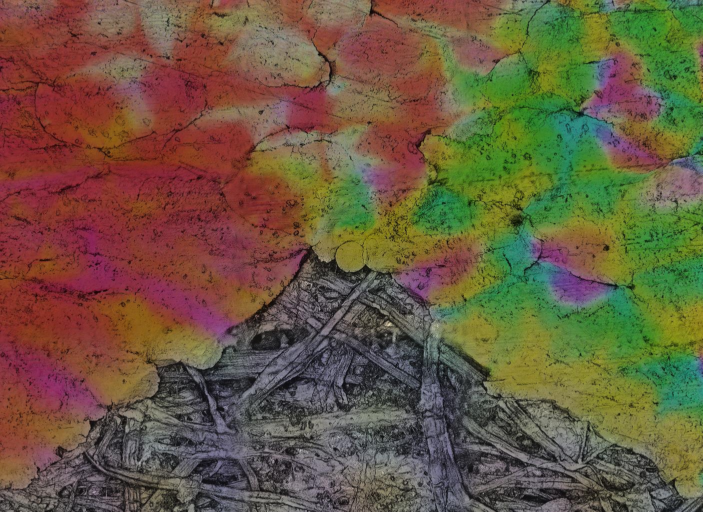
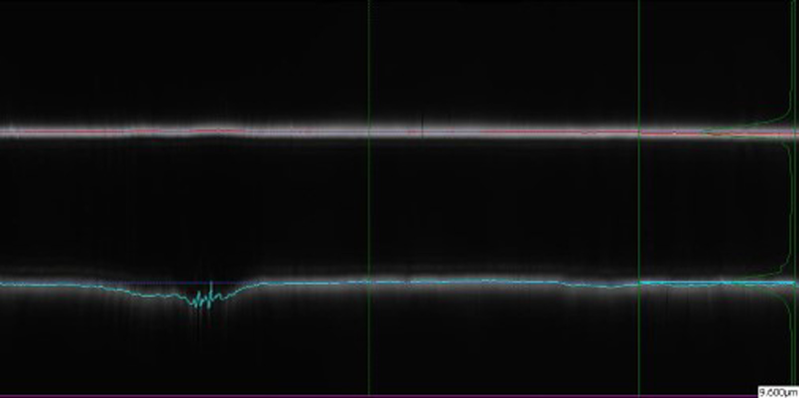
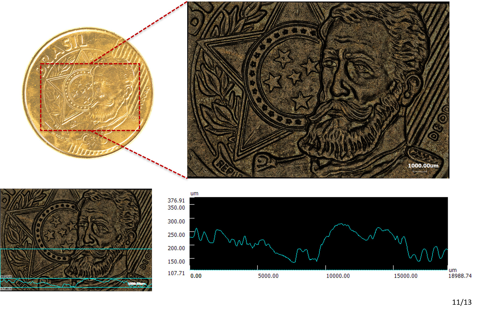
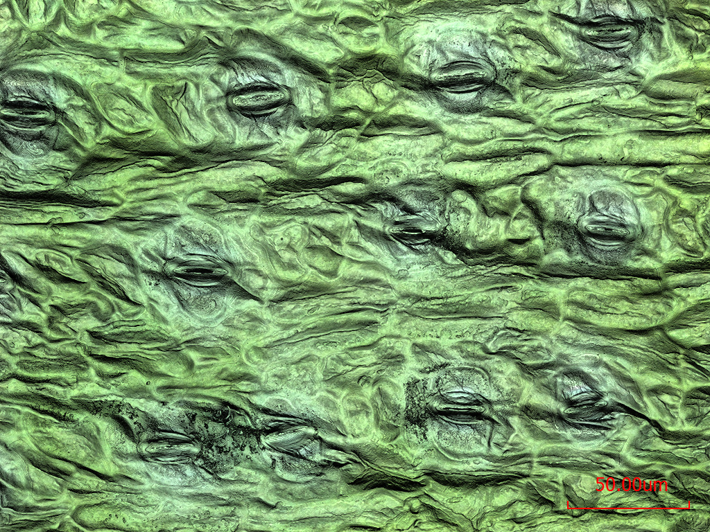
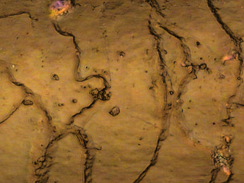
 Gallery