
LNNano is committed to the development of nanomaterials and new functional materials on a safe and sustainable basis in the direction of responsible innovation and Safe-by-Design. In this sense, we offer facilities and technical-scientific support to study the effects of nanomaterials on biological systems and the environment; aiming at a proactive assessment of potential hazards (toxicity) and risks involved during the development of these materials, including their functionalized derivatives, by-products and waste (life cycle). We support the generation of innovative products in Brazil in harmony with the protection of human, animal and environmental health.
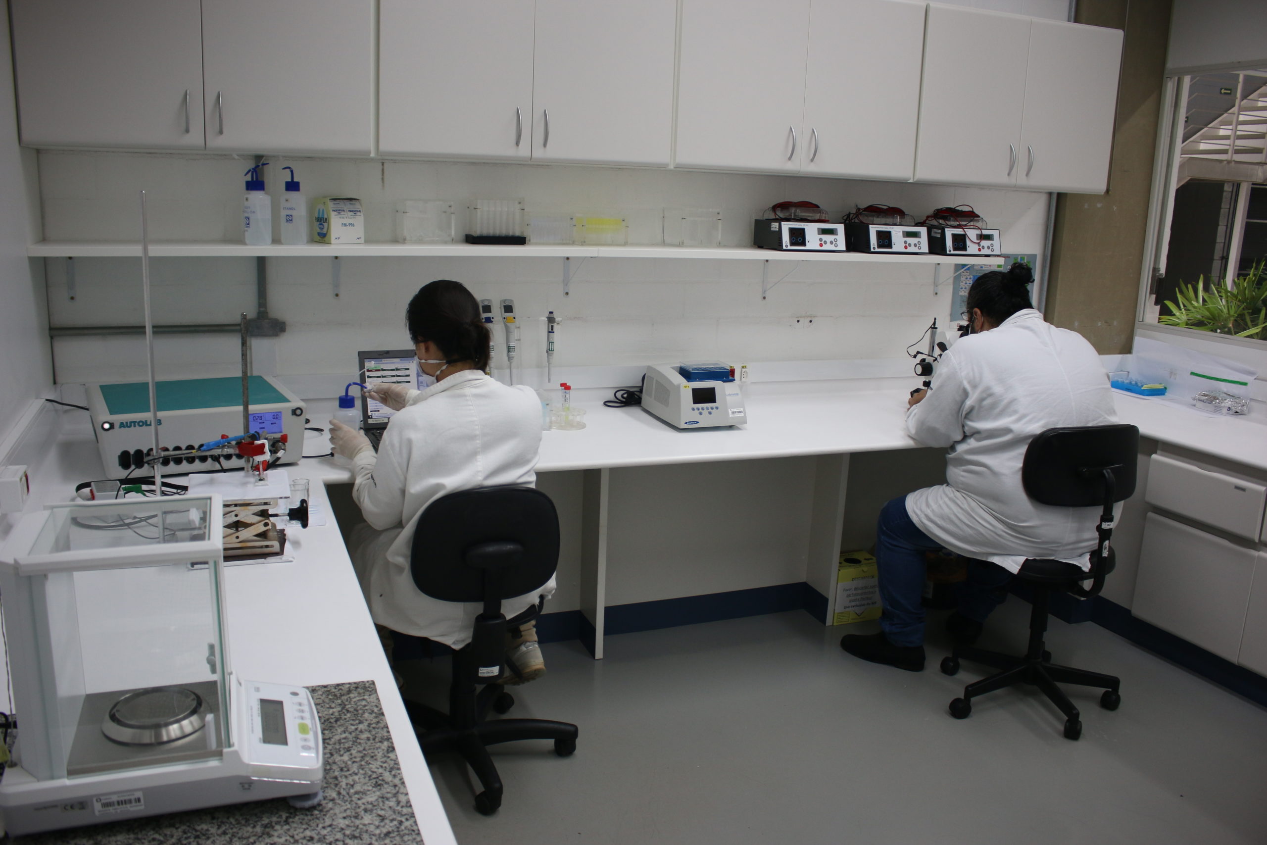
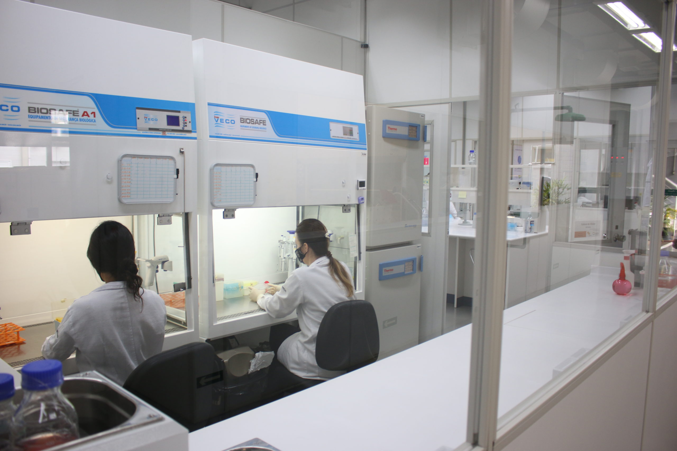
The Nanotoxicology and Nanosafety Facility develops and employs alternative methods to the use of animals in scientific experimentation – Principle 3Rs (Replacement, Reduction and Refinement). This facility has biosafety level NB-2 and provides personal protective equipment (PPE) for all its users; in addition to maintaining high standards of good laboratory practices (GLP) to guarantee the quality of research projects, results and services performed.


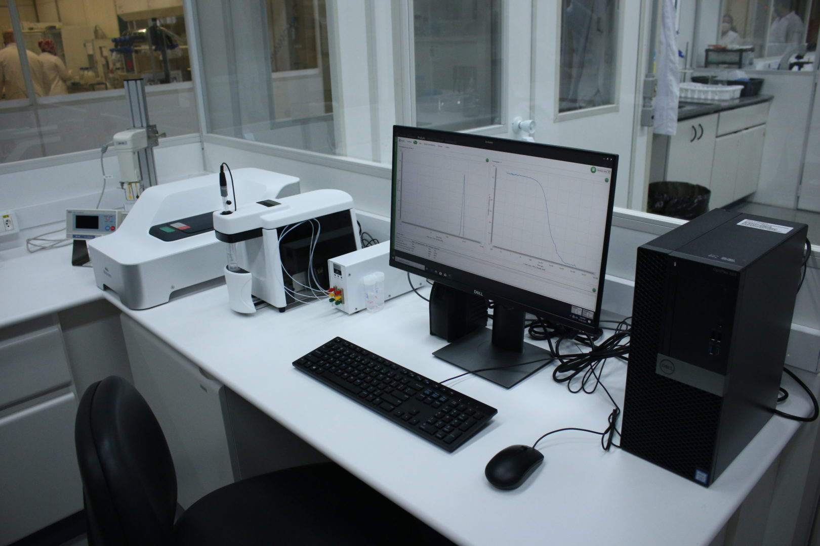
Infrastructure
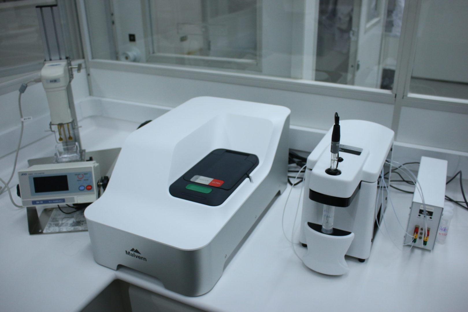
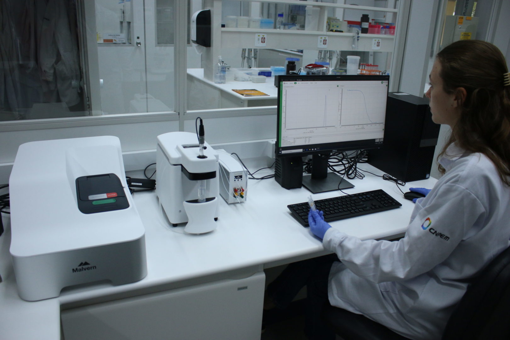
Dynamic and Electrophoretic Light Scattering (Zetasizer Ultra)
Dynamic and Electrophoretic Light Scattering (Zetasizer Ultra)
Sedimentation by Differential Centrifugation (CPS Disc Centrifuge)
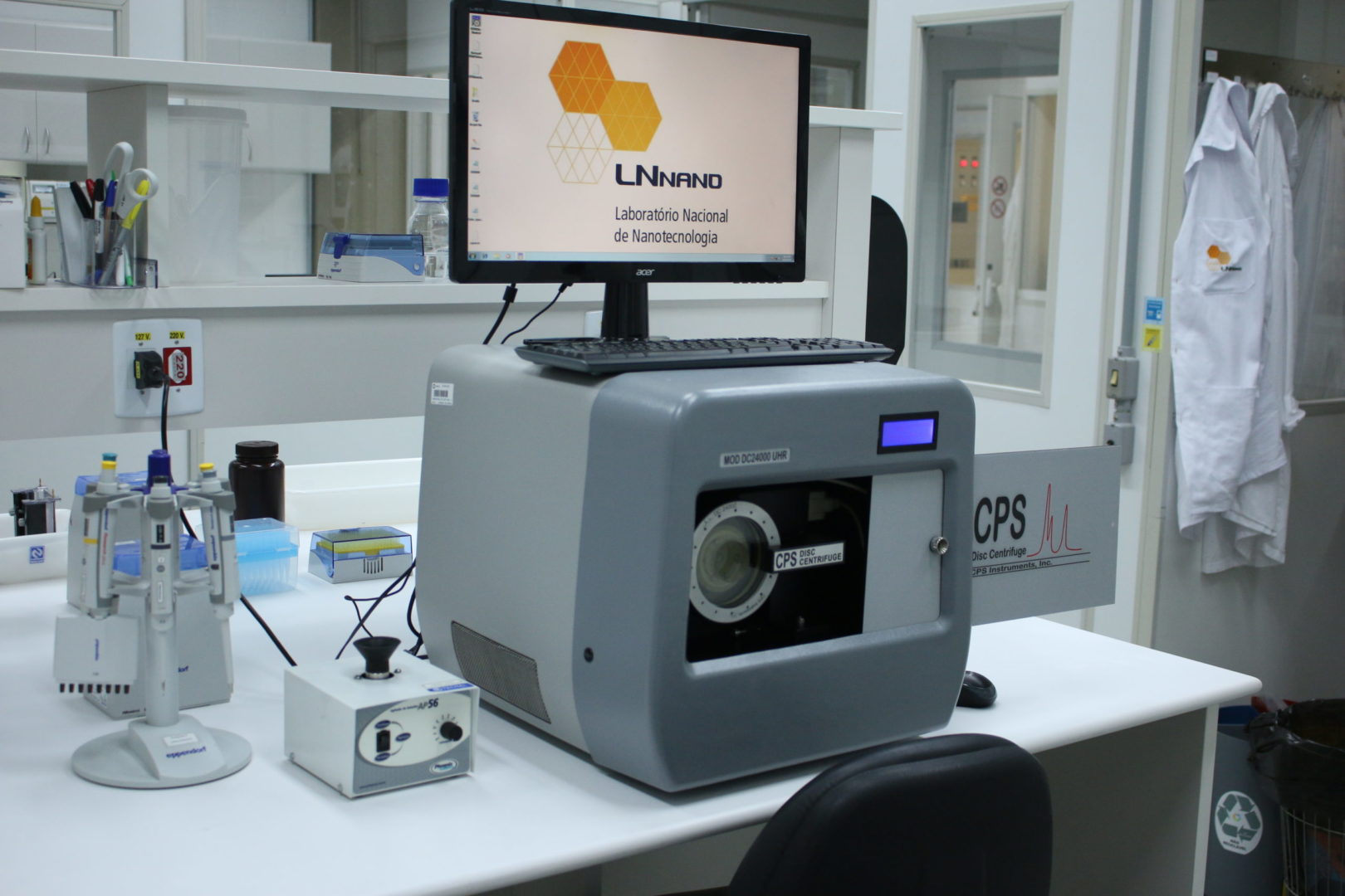

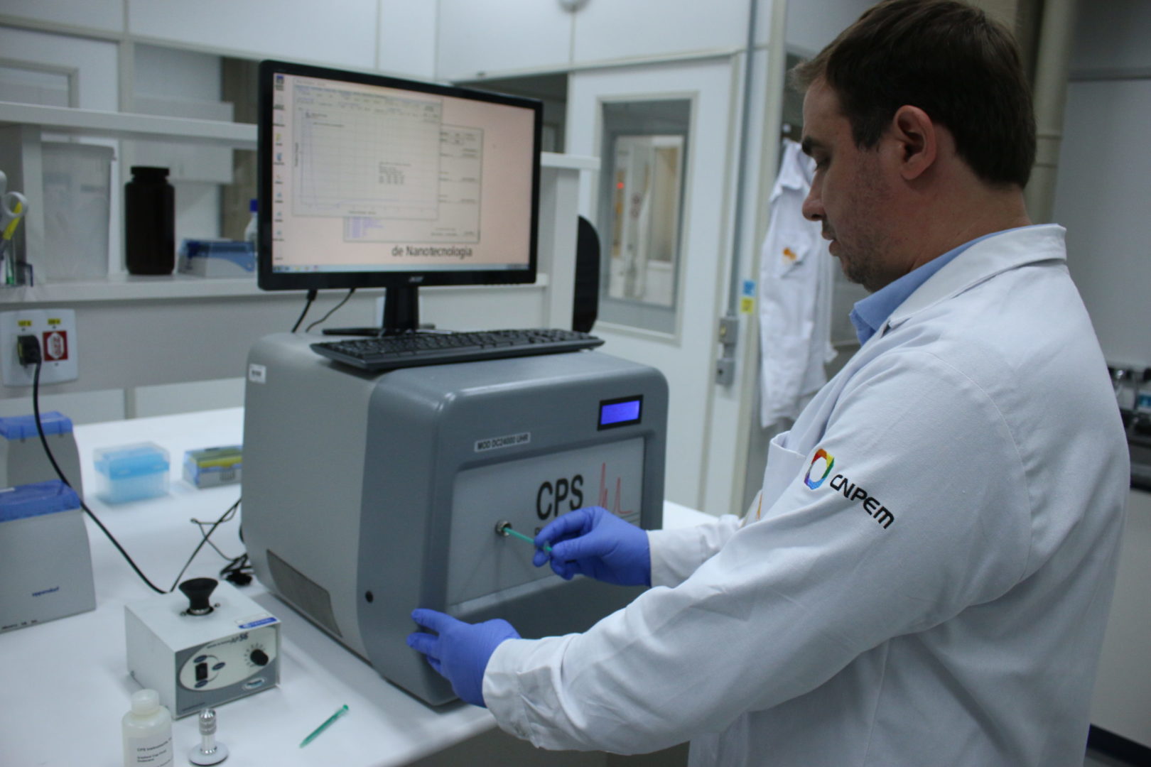
Sedimentation by Differential Centrifugation (CPS Disc Centrifuge)
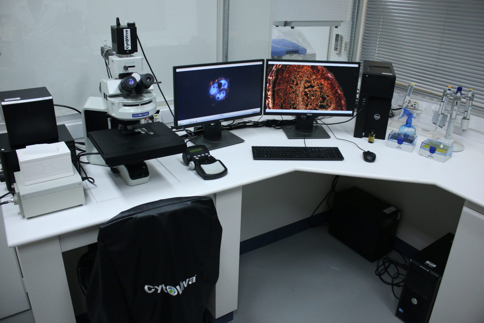
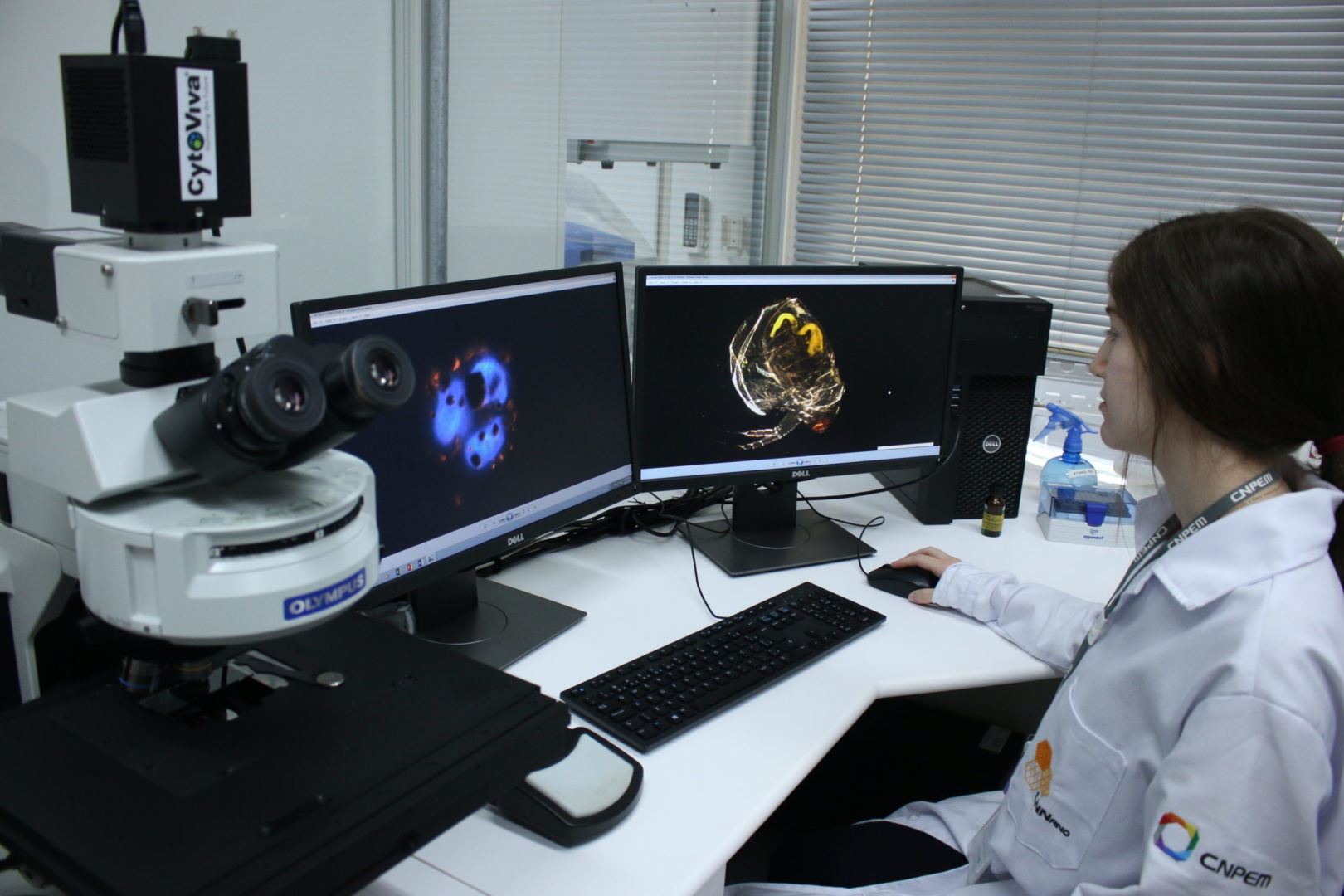
Darkfield Hyperspectral Microscopy (CytoViva)
Darkfield Hyperspectral Microscopy (CytoViva)
Toxicity – Zebrafish (ZFET)
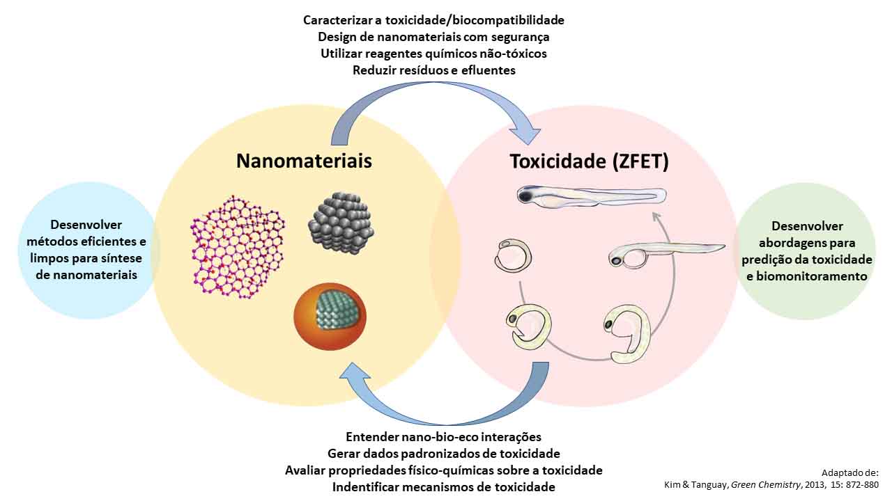


Toxicity – Zebrafish (ZFET)